Case 18
Case History
A 48-year-old woman presents for screening mammogram.
Physical Examination
• normal exam
Mammogram
Mass (Fig. 18–1)
• margin: circumscribed
• shape: oval
• density: equal density
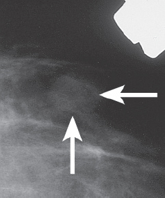
Figure 18–1. Left CC spot compression mammogram: In the outer breast there is an oval mass (arrows).
Ultrasound
Low Frequency
Frequency
• 7.5 MHz
Mass
• margin: well defined
• echogenicity: heterogeneous
• retrotumoral acoustic appearance: single edge shadowing
• shape: ellipsoid (Fig. 18–2)


Stay updated, free articles. Join our Telegram channel

Full access? Get Clinical Tree


