Case 18

 Clinical Presentation
A 37-year-old man with increasingly severe hypertension. A few months ago, the patient underwent lithotripsy for renal stones.
 Imaging Findings

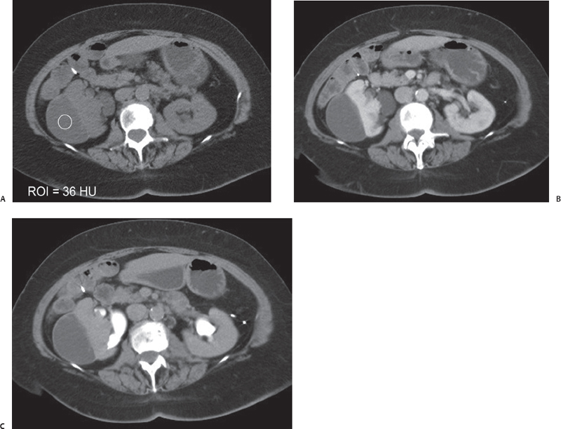
(A) Precontrast computed tomography (CT) image of the abdomen in the region of the kidneys shows a fluid collection (arrow) at the lateral aspect of the right kidney. The attenuation measurement of the fluid shown in the image reads 36 Hounsfield units, which is suggestive of a high protein content or altered blood. No perinephric stranding is seen. (B) Nephrographic phase contrast-enhanced CT image at the same level as Figure A shows no enhancement of the fluid collection. Renal perfusion is normal on both sides. The outline of the right kidney (arrowhead) is deformed by the collection, which appears to be under high pressure. (C) Excretory phase contrast-enhanced CT image at the same level as Figures A and B. Excreted contrast is seen in the collecting system (arrows). No dilatation of the collecting system is seen. No contrast is seen in the subcapsular fluid collection.
 Differential Diagnosis
• Subacute–chronic renal subcapsular hematoma:
Stay updated, free articles. Join our Telegram channel

Full access? Get Clinical Tree


