Variations of the Origin of the PICA
18.1 Case Description
18.1.1 Clinical Presentation
A 59-year-old man presented with a perimesencephalic subarachnoid hemorrhage. Computed tomography angiogram was negative for aneurysm, and digital subtraction angiography was obtained.
18.1.2 Radiologic Studies
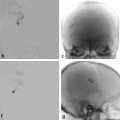
See ▶ Fig. 18.1.
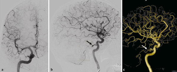
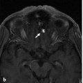
Fig. 18.1 Right ICA angiogram in lateral view in early (a) and late (b) arterial phases and 3D reconstruction (c) reveal a persistent trigeminal artery on the right side supplying the right PICA (arrows). The remainder of the intracranial arteries is unremarkable.
18.1.3 Diagnosis
Posterior inferior cerebellar artery (PICA) origin of a trigeminal artery. No cause for the hemorrhage was found.
18.2 Embryology and Anatomy
The PICA is the cerebellar artery with the most variations, which may range from unilateral or bilateral absence to being duplicated with different possible origins. The proximal PICA is the equivalent of a radiculopial artery that enlarged because of its annexed cerebellar territory via the primitive choroidal plexus of the fourth ventricle. The variations of the PICA origin follow the metameric arrangement also seen at the spinal cord level. Possible variations include the C3 origin from the vertebral artery (VA) or the ascending cervical artery; the C2 and C1 origins from the occipital artery via the embryonic proatlantal arteries (occipito-cerebellar variant); the ascending pharyngeal artery via the embryonic hypoglossal artery (pharyngo-cerebellar variant); and origin from various meningeal branches through the artery of the falx cerebelli. In exceedingly rare cases, there have also been reports of the PICA originating from the trigeminal artery.
The PICA is divided into five segments: the anterior medullary segment along the front of the medulla; the lateral medullary segment, which courses beside the medulla and extends to the origin of the glossopharyngeal, vagal, and accessory nerves; the tonsillomedullary segment, which courses around the caudal half of the cerebellar tonsil; the telovelotonsillar segment, which courses in the cleft between the tela choroidea and the inferior medullary velum rostrally and the superior pole of the cerebellar tonsil caudally; and finally, its cortical segment, with its course over the cerebellar surface.
Stay updated, free articles. Join our Telegram channel

Full access? Get Clinical Tree


