Case 2
Case History
A 39-year-old woman with new left breast lump. Left breast sonography is initially performed. As a result of the sonogram, a mammogram has been done.
Physical Examination
• left breast: 5 cm palpable lump in the upper inner quadrant
• right breast: normal exam
Mammogram
Mass (Fig. 2–1)
• margin: circumscribed
• shape: oval
• density: fat-containing
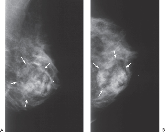
Figure 2–1. At the 9:00 position of the left breast, there is a well-defined oval fat-containing mass with heterogeneous density (arrows). (A). Left MLO mammogram. (B). Left CC mammogram.
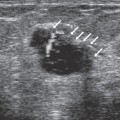
Ultrasound
Frequency
• 10 MHz
Mass
• margin: well defined
• echogenicity: heterogeneous


Stay updated, free articles. Join our Telegram channel

Full access? Get Clinical Tree


