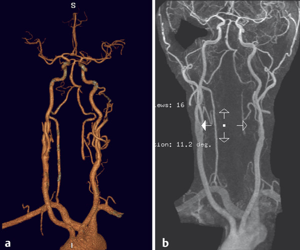
The Aberrant Subclavian Artery A 6-year-old boy with a history of aortic coarctation underwent a thoracic CTA for diagnostic workup. See ▶ Fig. 2.1. Fig. 2.1 (a,b) Coronal reformats and (c) 3D volume reconstruction of a thoracic CTA. The RCCA arises as a single vessel from the aortic arch (white arrow). The right subclavian artery has an aberrant origin and arises as a single trunk (dashed arrow) from the arch, distal to the left subclavian artery. The aortic coarctation is located between the left subclavian and the aberrant right subclavian (black arrow in c). Aberrant right subclavian artery with an aortic coarctation. Development of the aortic arch and great vessels results from the formation and selective involution of paired vascular arches connecting the embryonic aortic sac (ventral aorta) with paired dorsal aortae, as discussed in Case 1. Congenital aortic arch anomalies result from errors in the embryological development of the vascular arches and can be explained by abnormal persistence of arch segments that usually regress, involution of segments that usually remain, or both. A left aortic arch with an aberrant right subclavian artery (ARSA) is the most common congenital arch anomaly, occurring in approximately 0.4 to 2% of cases. During normal arch development, the segment of the dorsal aorta between the third and fourth arches obliterates, and the fourth vascular arches undergo asymmetric remodeling. On the right side, the fourth arch forms the most proximal portion of right subclavian artery, maintaining its connection to the definitive aortic arch through the brachiocephalic artery formed from the right horn of the aortic sac/ventral aorta. The subclavian artery more distally is formed from a segment of the right dorsal aortic arch and the right seventh intersegmental artery. The portion of the right dorsal aorta between the origin of the seventh intersegmental artery and the junction with the left dorsal aorta involutes. An ARSA occurs when there is abnormal obliteration of the right fourth vascular arch and the portion of the right dorsal aorta cranial to the seventh intersegmental artery. As a result, the right subclavian artery forms from the seventh intersegmental artery and the distal part of the right dorsal aorta. There is, therefore, interruption of the primitive arch segment between the right common carotid artery (RCCA) and right subclavian arteries, and the right subclavian artery arises directly off the aorta. As development proceeds, the origin of the right subclavian artery shifts cranially to lie just below the left subclavian origin. Therefore, the ARSA is the last branch of the aortic arch, arising distal to the left subclavian origin. Because its origin is derived from the right dorsal aorta, it must cross the midline posterior to the esophagus to perfuse the right upper extremity. Commonly, but not in all cases, a diverticular outpouching is present at the origin of the ARSA. Known as a diverticulum of Kommerell, this outpouching is thought to represent a remainder of the primitive right dorsal aorta (▶ Fig. 2.2). Fig. 2.2 MRA in anteroposterior (AP) view (a) and coronal MRA (b) demonstrate an aberrant right subclavian artery resulting from formation from the C7 segmental artery. Note the separate origins of the RCCA and LCCA in this case, rather than a common origin.
2.1 Case Description
2.1.1 Clinical Presentation
2.1.2 Radiologic Studies

2.1.3 Diagnosis
2.2 Embryology and Anatomy
2.3 Clinical Impact
Stay updated, free articles. Join our Telegram channel

Full access? Get Clinical Tree


