Diagnosis: Cecal volvulus
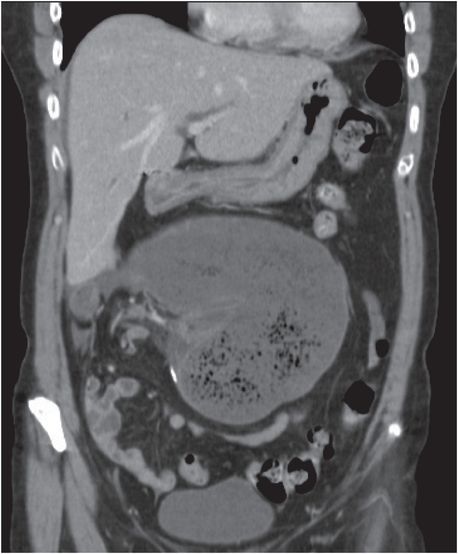
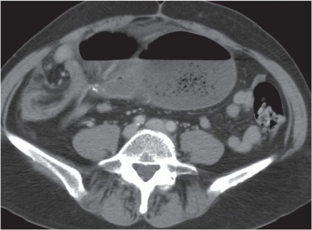
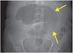
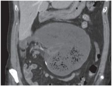
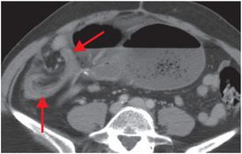
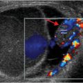
CT scout image (left image) shows a dilated, gas-filled viscus with haustral folds in the midline (yellow arrows). Coronal (middle image) and axial (right image) contrast-enhanced CT images show displacement of the dilated, fluid-filled cecum into the upper abdomen. There is an associated “whirl” sign (an area of swirling of the bowel and its mesentery, denoted by the red arrows) at the site of the twist, best seen on the axial image.
Discussion
Overview of cecal volvulus
Cecal volvulus accounts for 25–40% of all cases of colonic volvulus and 1% of intestinal obstructions.
Inadequate fixation of the right colon to the retroperitoneum, allowing increased mobility of the right colon, is the main underlying cause. The other requirement is restriction of the bowel at a fixed point, such as from scarring, adhesions, or an abdominal mass that serves as a fulcrum for rotation.
As in small bowel obstruction, patients may present with colicky abdominal pain, nausea, vomiting, and obstipation.
Imaging of cecal volvulus
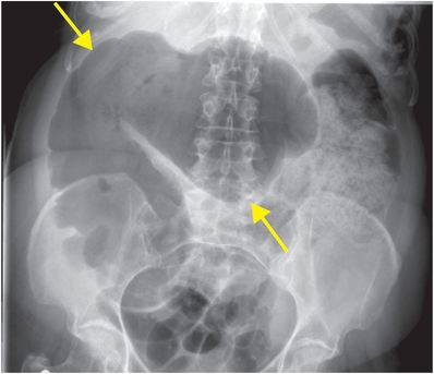
Cecal volvulus has a characteristic appearance on conventional radiography: a dilated loop of bowel with haustral markings, usually located in the left upper quadrant. The cecum, however, may be displaced anywhere in the abdomen. The loop may look like a coffee bean, similar to the radiographic sign that characterizes sigmoid volvulus.
Secondary signs of bowel ischemia should always be sought, including free air, portal venous gas, and cecal pneumatosis.
Small bowel proximal to the cecum is usually dilated.
After radiography, CT may be useful for diagnosing complications. CT findings include abnormal position of a markedly dilated cecum (typically the left upper quadrant, less commonly the right upper quadrant). Both bowel and mesentery are twisted, causing the “whirl” sign of swirling mesenteric vessels.
Contrast enema can confirm the diagnosis, characteristically showing a focal obstruction of the right colon with a beak-like narrowing at the site of twisting. Contrast enema is contraindicated in the setting of perforation, however.
Clinical synopsis
The patient underwent emergency exploratory laparotomy with ileocecectomy. Pathology revealed transmural chronic inflammation.
Self-assessment
|
|
|
|
Spectrum of cecal volvulus
Uncomplicated cecal volvulus
Uncomplicated cecal volvulus is volvulus without ischemia or infarction. Although the dilated loop in cecal volvulus is classically found in the left upper quadrant, the cecum can be located anywhere in the abdomen. Secondary signs of ischemia, including pneumatosis, portal venous gas, and pneumoperitoneum, are absent. Radiograph of the abdomen shows a dilated loop of bowel with haustral markings in the right upper quadrant (arrows).
Related posts:
 12 68-year-old man with left lower quadrant pain and hypotension
 32 32-year-old male complaining of chest pain after upper endoscopy
 28 30-year-old male presented with a palpable left testicular mass
 29 19-year-old male presented with acute onset right scrotal pain
 53 42-year-old female presenting with fever and back pain
 66 21-year-old male with quadriplegia after diving into a shallow pond. The patient struck his head against an embankment, with his head flexed, chin against chest
Stay updated, free articles. Join our Telegram channel

Full access? Get Clinical Tree


