Case 20
 Clinical Presentation
A 45-year-old woman who underwent computed tomography to evaluate for hematuria.
 Imaging Findings
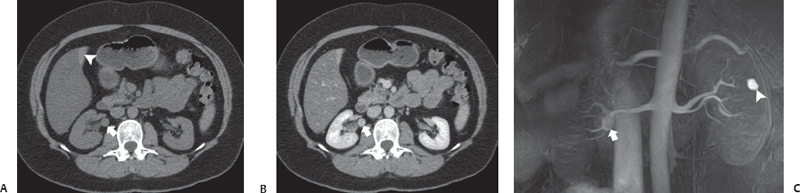
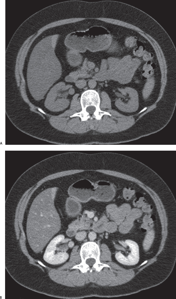
(A) Noncontrast computed tomography (CT) image of the abdomen in the region of the kidneys shows a small nodule (arrow) in the hilum of the right kidney. The visualized portions of both kidneys appear normal in size, shape, outline, and parenchymal thickness. The liver shows diffuse fatty infiltration with focal sparing in the gallbladder fossa (arrowhead). (B) Contrast-enhanced nephrographic phase CT image of the abdomen at the same level shows the nodule (arrow) to enhance uniformly in a fashion similar to that of the aorta. No filling defect is seen. No atherosclerotic changes are seen. (C) Noncontrast magnetic resonance (MR) angiogram performed afterward confirms the renal artery aneurysm (arrow). Incidental note is made of a simple cyst in the left kidney (arrowhead).
 Differential Diagnosis
• Right renal artery aneurysm: The location of the nodule in the right renal hilum with postcontrast enhancement following that of the aorta is typical.
• Retroperitoneal lymph node:
Stay updated, free articles. Join our Telegram channel

Full access? Get Clinical Tree


