Case 21
Case History
A 58-year-old woman presents with a new density on her screening mammogram.
Physical Examination
• right breast: inflamed obstructed follicle in the medial inferior breast
• left breast: normal exam
Mammogram
Mass (Fig. 21–1)
• margin: circumscribed
• shape: oval
• density: equal density
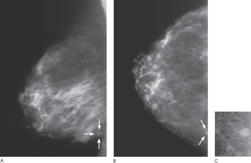
Figure 21–1. In the right medial inferior breast, there is a well-defined oval density (arrows). (A). Right MLO mammogram. (B). Right CC mammogram. (C). Right CC spot compression mammogram.
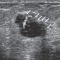
Ultrasound
Frequency
• 13.5 MHz
Mass


Stay updated, free articles. Join our Telegram channel

Full access? Get Clinical Tree


