Case 21
 Clinical Presentation
A 56-year-old man with a mass in the right lower quadrant.
 Imaging Findings
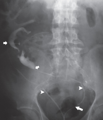
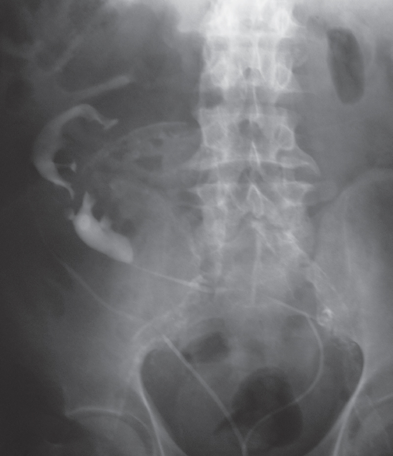
Retrograde pyelogram image shows that both ureters have been catheterized (arrowheads) and injected with contrast. There is no renal collecting system to the left of the midline. Both collecting systems are located to the right of the midline, lower than normal (at the level of the L4 and L5 vertebrae), and deformed. Both ureters arise from the collecting systems on the right of the midline (short arrows). The right ureter follows a tortuous course but inserts into the bladder to the right of the midline. The left ureter, after arising from the deformed kidney on the right of the midline, crosses the spine to the left to open into the urinary bladder on the left of the midline (long arrow). Both ureters and collecting systems are normal in caliber.
 Differential Diagnosis
• Crossed fused renal ectopia: The two kidneys are located on one side of the spine and fused. The left ureter is seen to be crossing the spine to its normal destination at the left upper angle of the trigone.
• Pancake kidney:
Stay updated, free articles. Join our Telegram channel

Full access? Get Clinical Tree


