Case 22

 Clinical Presentation
A 38-year-old woman with urinary tract infections. An intravenous pyelogram was performed.
 Imaging Findings

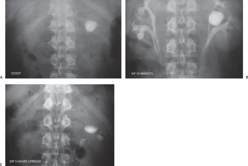
(A) Scout image of an intravenous pyelogram (IVP) in the kidney area shows a round calcific density (arrow) overlying the left renal area. (B) An IVP radiograph of the renal area obtained at 10 minutes after the injection of contrast shows bilateral duplex collecting systems (arrows). The previously seen calcific density (arrowhead) is seen to lie in the vicinity of the left upper pole calix and shows increased density, suggesting some entry of excreted contrast. (C) Upright delayed radiograph shows some residual contrast (arrows) in the left collecting system. This is of no clinical significance. However, the previously seen calcific density has a different shape and now shows a fluid level at its upper margin (arrowhead).
 Differential Diagnosis
• Caliceal diverticulum with milk of calcium urine:
Stay updated, free articles. Join our Telegram channel

Full access? Get Clinical Tree


