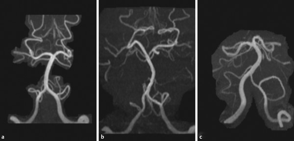
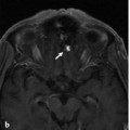
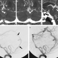
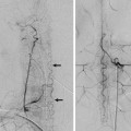
Fig. 22.1 Right vertebral artery angiogram in 3D reconstruction demonstrates a broad-based basilar tip aneurysm that is arising from the caudal P1 in the setting of asymmetric fusion of the basilar tip. Note that a single bithalamic perforator (arrow) to the thalamus (so-called artery of Percheron) is present, arising from the cranial P1.

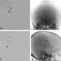
Fig. 22.2 Right vertebral artery angiograms in the working projection before (a) and after (b) coiling. In this scenario, the caudal P1 is at higher risk of occlusion, given its broad communication with the aneurysm. A working projection should, however, also ensure that the origin of the Percheron artery is well visualized.
22.1.3 Diagnosis
Saccular aneurysm in the presence of an asymmetric fusion of the basilar tip.
22.2 Embryology and Anatomy
The termination of the basilar artery can be subdivided into three distinct types, depending on the time when the caudal portions of the distal internal carotid artery (ICA) merge with the paired ventral longitudinal neural arteries: a cranial symmetric fusion type, a caudal symmetric fusion type, or an asymmetric fusion type. As pointed out in the previous case, formation of the basilar artery occurs via a bidirectional fusion process. The fusion process cranial to the trigeminal point incorporates merging of both caudal divisions of the embryonic ICAs with the anterior longitudinal neural arteries. If the fusion occurs early, it will be completed from its most cranial point onward and will lead to fusion of both the right and the left caudal division of the ICA to the level of the pontomesencephalic sulcus, leading to an adult T-shape of the basilar tip (▶ Fig. 22.3). This fusion type constitutes the most “mature” or complete fusion and is referred to as a “cranial” fusion type. If the fusion of both limbs occurs later, it will be below the pontomesencephalic sulcus, and a V-shape of the basilar tip will ensue; this is referred to as a “caudal” fusion type. The third type of fusion is when merging is asymmetrical, with one caudal division merging with the anterior longitudinal neural arteries earlier (more cranially) than the other caudal division of the ICA. This disposition is called an asymmetric fusion.
