Case 23
 Clinical Presentation
A 51-year-old man with right flank pain. Stone protocol computed tomography was performed.
 Imaging Findings

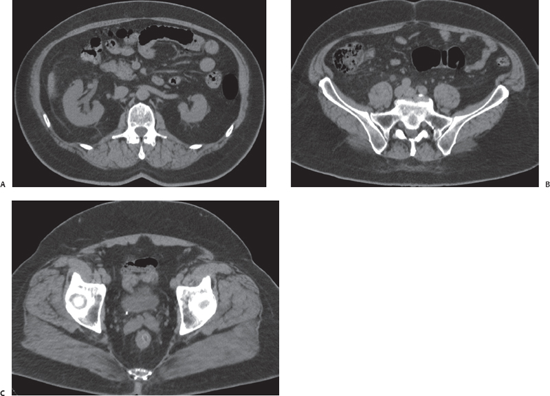
(A) Noncontrast computed tomography (CT) image at the level of the kidneys shows that the right kidney is mildly enlarged compared with the left. There is fullness of the collecting system on the right (arrow). There is mild right perinephric stranding (arrowhead). (B) Noncontrast CT image at the midureteral level shows that the right ureter (arrow) is mildly dilated. (C) Noncontrast CT image at the level of the ureterovesical junctions (UVJs). There is a stone (arrow) in the lower end of the right ureter.
 Differential Diagnosis
• Right ureteric calculus: A calcific density in a ureter with dilatation of the part of the ureter and collecting system proximal to it is characteristic. The perinephric stranding shows that the obstruction has been present for at least a few hours.
• Phlebolith in the pelvic vein:
Stay updated, free articles. Join our Telegram channel

Full access? Get Clinical Tree


