Case 24
 Clinical Presentation
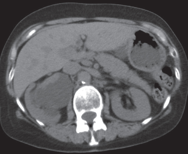
A 72-year-old man with a history of smoking and recent painless hematuria.
 Imaging Findings
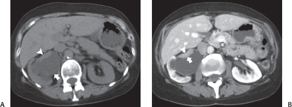
(A) Precontrast computed tomography (CT) image at the level of the kidneys shows dilatation of the right renal collecting system (arrow). There is a focal plaquelike thickening (arrowhead) on the anterior wall of the right renal pelvis. (B) Contrast-enhanced CT image at the same level shows enhancement of the plaque (arrow) on the anterior wall of the right renal pelvis. There is retraction (arrowhead) of the wall of the renal pelvis underlying the plaque. Delayed enhancement of the right renal parenchyma is due to obstruction.
 Differential Diagnosis
• Transitional cell carcinoma (TCC) arising from the right renal pelvis: Focal thickening of the wall of the renal pelvis should be considered malignant unless proven otherwise. TCC is the most common neoplasm arising from the wall of the renal pelvis.
• Pyelitis cystica profunda:
Stay updated, free articles. Join our Telegram channel

Full access? Get Clinical Tree


