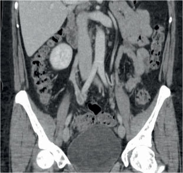
Diagnosis: Gonadal (ovarian) vein thrombosis
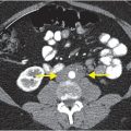
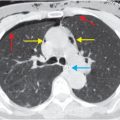
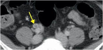
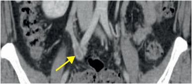
Contrast-enhanced axial (left image) and coronal (right image) CT demonstrate a non-occlusive, linear filling defect in the right gonadal vein (arrows).
Discussion
Overview of ovarian vein thrombosis
Ovarian vein thrombosis is a rare but serious condition that may occur in the postpartum period or in patients with PID, malignancy, or recent pelvic surgery.
Patients with ovarian vein thrombosis may be asymptomatic or present with vague abdominal pain and fever; if the condition is untreated, pulmonary embolism, sepsis, or even death may result.
Mainstays of treatment include intravenous antibiotics and therapeutic anticoagulation. In some cases, surgery may be indicated to treat the underlying condition.
Ovarian veins arise from the broad ligament and extend superiorly in the retroperitoneum, anterior to the psoas muscle. The right gonadal vein enters the IVC directly, whereas the left gonadal vein drains into the left renal vein.
Ovarian vein thrombosis can be detected using ultrasound or cross-sectional imaging modalities such as CT or MRI.
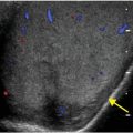
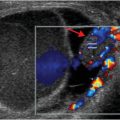
Ultrasound is often used as the first-line imaging test, given its wide availability – however, it has lower sensitivity and specificity than MRI or CT. The typical ultrasound appearance is an enlarged vein with an intraluminal, echogenic filling defect.
On contrast-enhanced CT, a gonadal vein thrombus may appear as a central filling defect within the opacified vein. When occluded, the thrombosed vein may appear as a non-enhancing tubular structure in the retroperitoneum. In some cases, the vein may be enlarged with surrounding inflammatory fat stranding. Multiplanar imaging helps distinguish the ovarian vein from other tubular structures such as the appendix, ureter, and bowel.
MRI findings include a variable-signal-intensity filling defect on T1-weighted contrast-enhanced images and an intermediate to high signal thrombus with a dark peripheral rim (hemosiderin) on T2-weighted images.
Stay updated, free articles. Join our Telegram channel

Full access? Get Clinical Tree


