Case 25
 Clinical Presentation
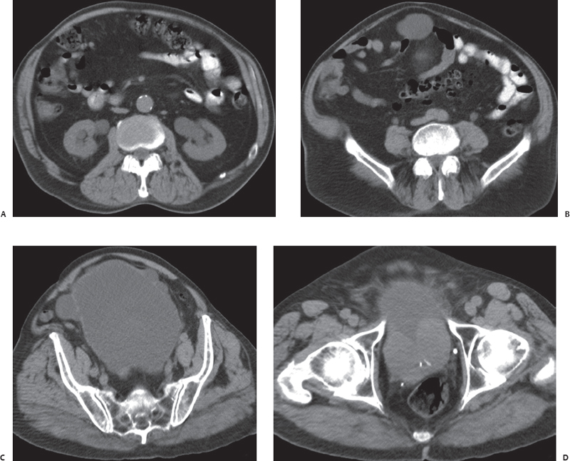
A 68-year-old man with deteriorating renal function. Computed tomography was performed for further evaluation of hydronephrosis seen on ultrasound.
 Imaging Findings
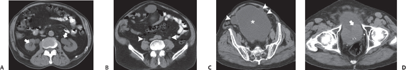
(A) Noncontrast computed tomography (CT) image at the level of the kidneys shows dilatation of the collecting systems (arrows) of both kidneys. (B) Noncontrast CT image at the midureteral level shows dilatation of both ureters (arrowheads). (C) Noncontrast CT image at the level of the urinary bladder shows dilatation of the urinary bladder (asterisk) with multiple bladder diverticula (arrowheads). (D) Noncontrast CT image at the level of the prostate shows marked prostatic enlargement (arrow).
 Differential Diagnosis
• Bladder outlet obstruction caused by an enlarged prostate: Bilateral hydronephrosis, bilateral hydroureter, and abnormalities of the urinary bladder wall, along with bladder diverticula, are characteristic features in patients with problems of bladder emptying. An enlarged prostate is the cause.
• Abnormal bladder emptying caused by detrusor sphincter dyssynergia:
Stay updated, free articles. Join our Telegram channel

Full access? Get Clinical Tree


