Case 26
Case History
A 69-year-old woman presents for screening mammogram.
Physical Examination
• normal exam
Mammogram
Mass (Fig. 26–1)
• margin: indistinct
• shape: oval
• density: equal density
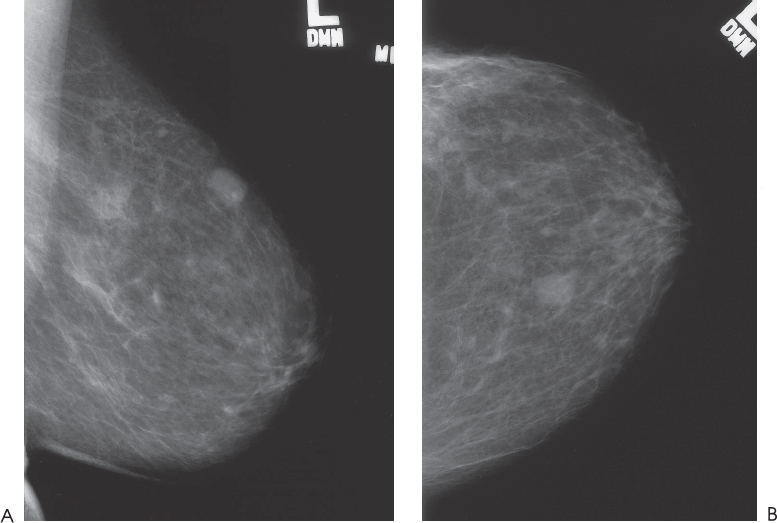
Figure 26–1. In the 1:00 position of the left breast, there is an oval mass that is new since the patient’s previous screening mammogram. The margins are well defined in the MLO view but indistinct in the CC view. (A). Left MLO mammogram. (B). Left CC mammogram.
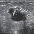
Ultrasound
Frequency
• 13 MHz
Mass
• margin: well defined, microlobulated
• echogenicity: hypoechoic
• retrotumoral acoustic appearance: bilateral edge shadowing


Stay updated, free articles. Join our Telegram channel

Full access? Get Clinical Tree


