Case 26

 Clinical Presentation
A 42-year-old man with a recent cardiac valve replacement developed hematuria and urinary stream interruption. Computed tomography urography was performed for further evaluation. The upper urinary tract was normal bilaterally.
 Imaging Findings

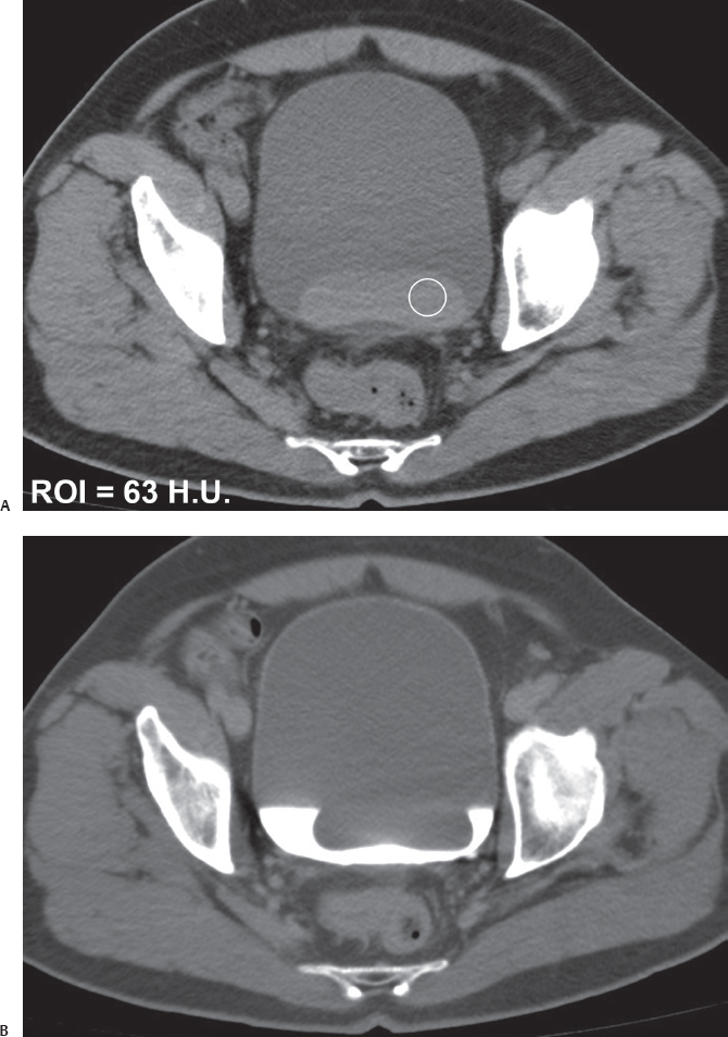
(A) Precontrast computed tomography (CT) image at the level of the urinary bladder shows a high-attenuation mass (arrow) surrounded by low-attenuation urine (asterisk) in the urinary bladder lumen. The mass has a smooth outline. Its attenuation value in the region of interest encompassed by the circle has been measured at 63 Hounsfield units. No retraction of the urinary bladder wall is noted in the areas of contact with the mass (arrowheads). No pelvic lymphadenopathy is seen in the image provided. (B) Excretory phase CT image at the same level as Figure A shows the mass (arrow) as a filling defect floating in the contrast-opacified urine (asterisk).
 Differential Diagnosis
• Blood clot in the urinary bladder:
Stay updated, free articles. Join our Telegram channel

Full access? Get Clinical Tree


