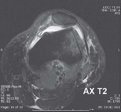
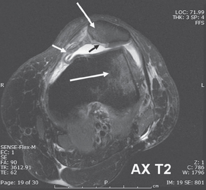
Case 28 A 30-year-old woman presents with severe knee pain. Axial T2-weighted image through the patellofemoral joint displays contusions over the periphery of the lateral condyle and medial patella (long white arrows). There is disruption of the medial patellofemoral retinaculum (short white arrow), with a chondral defect centered over the patellar apex (short black arrow

 Clinical Presentation
 Imaging Findings

![]()
Stay updated, free articles. Join our Telegram channel

Full access? Get Clinical Tree


