Case 28
 Clinical Presentation
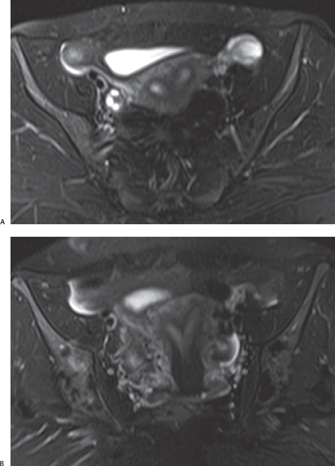
A 36-year-old woman who underwent pelvic magnetic resonance imaging for repeated miscarriages.
 Imaging Findings
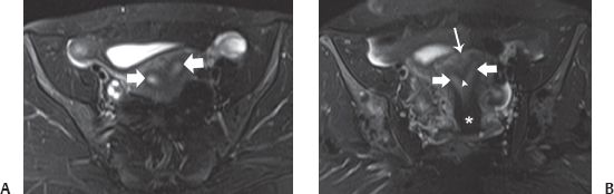
(A) Transverse fat-saturated T2-weighted image through the pelvis shows two separate endometrial canals (arrows) separated by myometrium. The thickness of the endometrium appears normal. (B) Coronal fat-saturated T2-weighted image through the pelvis confirms the presence of two endometrial canals (short arrows). They are separated by a myometrial septum that is incomplete (arrowhead). The fundus (long arrow) is smooth without any notch. At a more inferior level, only one cervix (asterisk) is identified.
 Differential Diagnosis
• Septate uterus: A double endometrial canal is always abnormal. Because the myometrium is not split into two horns, the fundus of the uterus is convex, and the septum does not extend through the entire length of the endometrial canal. This appearance is typical of a septate uterus.
• Arcuate uterus:
Stay updated, free articles. Join our Telegram channel

Full access? Get Clinical Tree


