Case 29
 Clinical Presentation
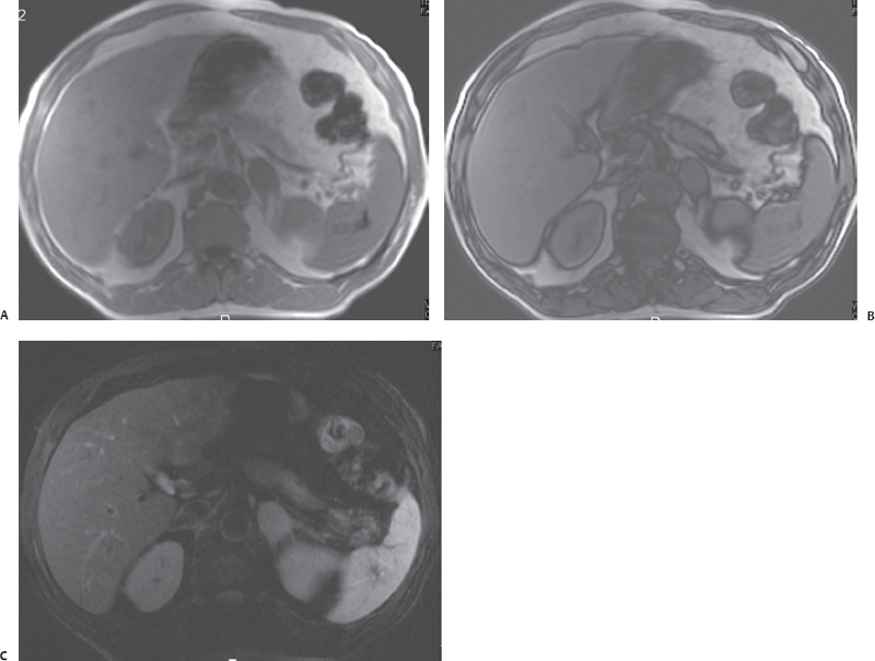
A 52-year-old man with bronchogenic carcinoma. Magnetic resonance imaging was performed for evaluation of an adrenal nodule seen on computed tomography.
 Imaging Findings
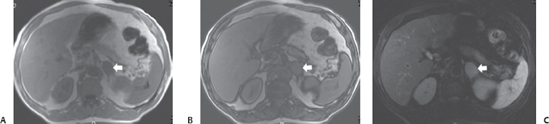
(A) In-phase T1-weighted magnetic resonance imaging (MRI) of the abdomen at the level of the adrenal gland shows a nodule (arrow) in the left adrenal gland. It is of intermediate signal intensity. (B) Opposed-phase T1-weighted MRI of the abdomen at the same level as Figure A shows that the adrenal nodule (arrow) maintains its signal. No loss of signal has been demonstrated. (C) Fat-saturated T2-weighted MRI of the abdomen at the same level as Figures A and B shows that the left adrenal nodule (arrow) has homogeneous intermediate signal intensity. It does not show fluid.
 Differential Diagnosis
• Adrenal metastasis:
Stay updated, free articles. Join our Telegram channel

Full access? Get Clinical Tree


