29 Filum Terminale Lipoma
29.1 Case Presentation
29.1.1 History
A 9-year-old male patient presents with a history of nonspecific back pain.
29.2 Imaging Analysis
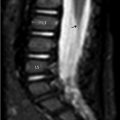
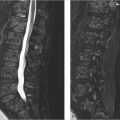
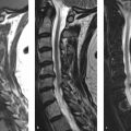
Lumbosacral spine MRI from a 9-year-old male patient with nonspecific back pain was performed. Sagittal T1-weighted (T1w; ▶ Fig. 29.1a), sagittal short tau inversion recovery (STIR; ▶ Fig. 29.1b), axial T1w (▶ Fig. 29.1c), and axial T2-weighted (T2w) with fat suppression (▶ Fig. 29.1d) images. An intrathecal linear hyperintensity is seen in T1w sagittal and axial images (arrows in ▶ Fig. 29.1a,c), in close relationship with the filum terminale; its high T1w signal intensity is suppressed in the series with fat saturation (arrows in ▶ Fig. 29.1b,d). Note the normal morphology and position of conus medullaris (asterisk in ▶ Fig. 29.1b) with tip located at the superior end plate of L2 level (arrowhead in ▶ Fig. 29.1b).

29.3 Differential Diagnosis
Related posts:
Stay updated, free articles. Join our Telegram channel

Full access? Get Clinical Tree


