32 Low-Lying Conus
32.1 Case Presentation
A 9-month-old, former 30-week premature male patient, presents with a history of mosaic tetrasomy 22q (cat eye syndrome). No spine-related symptoms or signs were found at physical examination (PE).
32.2 Imaging Analysis
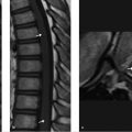
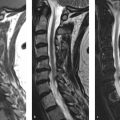
Sagittal short tau inversion recovery (STIR) image of the lumbar spine from a 9-month-old infant (▶ Fig. 32.1). The conus medullaris tip is located at mid L3 level (arrow), without any other associated cord, thecal sac, bone, or filum abnormality.

32.3 Differential Diagnosis
Low-lying conus medullaris:
It refers to a low position of a normal-appearing conus medullaris with respect to the vertebral level.
It is usually located between the T12–L1 and L1–L2 disk level; however, in 6.4% of population it can be found between the upper and middle third of L2. 1
Tethered spinal cord:
A low-lying conus medullaris pulled down by a thick filum terminale.
The conus medullaris is located at or below the inferior end plate of L2 and is attached to a thickened filum or filum lipoma.
In 25% of cases, there is central canal dilatation. 2
Open or closed spinal dysraphism:
This is a wide spectrum of malformations including spinal lipoma, myelomeningocele, meningocele, dermoid, and dermal sinus tract.
32.4 Imaging Pearls
Related posts:

Stay updated, free articles. Join our Telegram channel

Full access? Get Clinical Tree


