31 Prominent Central Canal
31.1 Case Presentation
A 16-year-old female patient with history of a motor vehicular collision presents with complaint of neck pain and thoracic region back pain.
31.2 Imaging Analysis
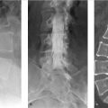
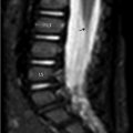
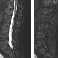
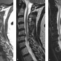
Spine MRI from a 16-year-old female patient with sagittal T2-wighted (T2w; ▶ Fig. 31.1a), sagittal T1-weighted (T1w; ▶ Fig. 31.1b), and axial T2w (▶ Fig. 31.1c) images. A slightly enlarged central canal is noticed (arrows); however, there are no additional abnormalities of the spinal cord in course, overall diameter, or surrounding parenchymal signal intensity.

31.3 Differential Diagnosis
Prominent central canal:
This refers to a slightly expanded central canal filled with cerebrospinal fluid (CSF) without any spinal cord signal abnormality or enhancement.
Cystic spinal cord neoplasm:
The imaging hallmarks are cord signal abnormality, mass effect, contrast enhancement, and associated with neurological symptoms. 1
Syringohydromyelia:
Cystic dilatation of the central canal that could be isolated or associated with congenital anomalies in up to 30% of cases. 1
The entire spine MRI should be performed to rule out low-lying cerebellar tonsils (Chiari I malformation) as well as low-lying conus with or without fatty filum (tethered cord).
Myelomalacia:
Cord atrophy secondary to previous vascular, traumatic, or other injury.
Related posts:
Stay updated, free articles. Join our Telegram channel

Full access? Get Clinical Tree


