Bladder Calculi
Nephroblastoma (Wilms’ Tumor)
Nephrocalcinosis
Nephrolithiasis (Renal Calculi)
Ovarian Dermoid Cysts
Other Ovarian Lesions
Pheochromocytoma
Renal Cell Carcinoma
Uterine Fibroma (Leiomyoma)
Portions of the genitourinary system are readily visible on plain film radiographs, which may aid in the localization of abnormal densities. This is a common site for concretions and some calcifying masses. Table 29-1 lists the common appearance of the genitourinary system and some of the abnormal findings that may be visible on plain film.
| Organ | Plain film visibility | Plain film location | Retroperitoneal versus intraperitoneal | Fixed versus mobile | Appearance specifics | Plain film abnormalities |
|---|---|---|---|---|---|---|
| Kidneys | Surrounding pericapsular fat | T12 to L2 level, right slightly lower than left | Retroperitoneal | Fixed; some translation with upright posture or inspiration | Height = 2 to 2.5 vertebral bodies plus intervertebral disc (IVD); long axis oriented superomedial to inferolateral | Lobulated contour; enlargement; ectopic; abnormal axis; nephrolith |
| Adrenals | None | Adjacent to body margins of T12 or L1 | Retroperitoneal | Fixed | None | Calcifications (masslike or cystic) |
| Ureters | None | Along anterior aspect of psoas muscle to posterosuperior bladder | Intraperitoneal | Fixed | None | Nephrolith |
| Bladder | Domed water density | Midline in pelvic bowl | Intraperitoneal | Fixed | Inferior margin less than 1 cm above pubic symphysis; superior margin may be indented by uterus | May appear as mass, may become distended (e.g., neurogenic bladder); superiorly displaced by enlarged prostate |
| Prostate | None | Midline at pubic symphysis | Intraperitoneal | Fixed | None | Calcifications |
| Uterus | None | Midline, pelvic bowl | Intraperitoneal | Fixed | None | Calcifications (fibroids) |
| Ovaries | None | Variable | Intraperitoneal | Somewhat mobile | None | Calcifications (dermoid cyst) |
Angiomyolipoma (Hamartoma)
BACKGROUND
Angiomyolipoma (AML) is the most common benign renal neoplasm diagnosed radiologically. It is a hamartomatous tumor containing varying amounts of fat, smooth muscle, and vascular tissues, and it may range in size from a few millimeters to more than 20 cm. 21 AMLs are found in young adults to elderly patients and are four and a half times more common in women. 10,21 Multiple, bilateral, small AMLs that may be symptomatic are found in up to 80% of patients with tuberous sclerosis, an uncommon heritable condition. 10,13,21 However, most angiomyolipomas arise sporadically.
IMAGING FINDINGS
The radiolucent fatty tissue of these tumors is seldom appreciated on plain film radiographs. The AML may be revealed by its mass effect after contrast injection on an intravenous pyelogram but cannot be differentiated from other masses. Attenuation of the collecting system with displacement and focal dilation of the calyces may be visible. 21
A renal tumor containing fat density tissue is essentially diagnostic for AML. Unenhanced computed tomography (CT) scans, use of small areas for attenuation measurements, and use of thin sections may be necessary to detect small amounts of fat. 3,12,21
Sonography characteristically shows a very hyperechoic lesion compared with adjacent renal parenchyma. * The high fat content, heterogeneous cellular architecture, and presence of multiple vessels all contribute to the marked echogenicity, but still this lesion cannot accurately be separated from renal cell carcinoma on ultrasound; therefore CT is necessary. 14,21
CLINICAL COMMENTS
Angiomyolipomas generally are asymptomatic and usually are identified during evaluation for unrelated genitourinary conditions. Flank pain, hematuria, and palpable mass are classic symptoms and usually are seen in otherwise healthy patients with isolated, unilateral lesions. 21 Parenchymal, subcapsular, or perirenal hemorrhage may lead to hypotension. 21
Embolization or partial excision is usual for large or symptomatic lesions. Overall, treatment for AMLs is usually conservative with follow-up alone being sufficient for most small, asymptomatic tumors. 10,21
• Angiomyolipoma is the most common radiologically diagnosed benign renal neoplasm.
• Classic symptoms, when present, are flank pain, hematuria, and palpable mass.
• Fatty attenuation seen on computed tomography is virtually diagnostic for angiomyolipoma.
Bladder Calculi
BACKGROUND
Ninety-eight percent of bladder calculi identified in the United States occur in elderly men. 16 The instigating factor in their formation usually is urinary retention with high residual urine. Superimposed infections add to susceptibility. The presence of bladder stones is associated with an increased incidence of carcinoma. 16
IMAGING FINDINGS
Bladder calculi often are nonopaque or only faintly opaque, allowing them to be frequently overlooked. Fecal material and gas in the rectosigmoid colon and the sacrum itself also overlie the area and may further obscure them. Bladder calculi usually are located centrally in the pelvis (Fig. 29-1). Those that are found more laterally may lie in a bladder diverticulum. 16
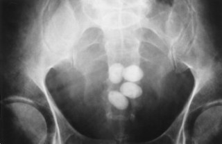 |
| FIG. 29-1 Multiple bladder calculi. Four oval radiopaque bladder stones are visible centrally in the pelvis. Most bladder calculi are round or oval but they may be amorphous, laminated, or even spiculated. |
Bladder calculi may abrade and irritate the mucosa and predispose to infection. Bladder infections are more difficult to treat when calculi are present. Ureteral or bladder outlet obstruction may also be present. 16 Approximately 15% of patients with gout, as well as hyperuricemic patients, produce uric acid stones. 16
Standard treatment of bladder stones consists of endoscopic visualization and fragmentation by electrohydraulic probe. 31 Massive calculi may require open surgery. 31
• Urinary retention with high residual urine is a common cause for formation.
• It may be difficult to detect on plain radiographs.
• Bladder calculi usually lie centrally in the pelvis on plain radiographs.
• The incidence of carcinoma of the bladder increases in the presence of stones.
Nephroblastoma (Wilms’ Tumor)
BACKGROUND
Nephroblastoma (Wilms’ tumor) is the most common abdominal malignancy in childhood. 8,13 Overall, leukemia-lymphoma, brain tumors (astrocytoma and medulloblastoma), and neuroblastoma have a higher incidence in pediatric populations when other sites of the body are also considered. 19 The highest incidence occurs in 3- and 4-year-old individuals, with 80% of Wilms’ tumors occurring in individuals 1 to 5 years old. 2,5,19
IMAGING FINDINGS
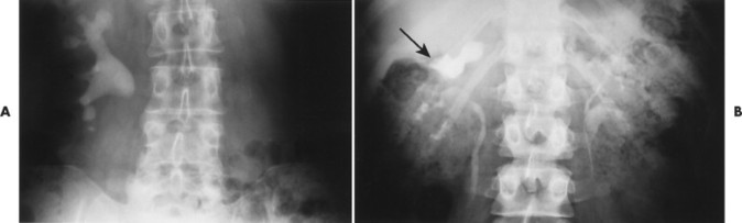
Wilms’ tumor appears radiographically as a complex renal mass (Fig. 29-2). Approximately 5% to 10% of these lesions show areas of calcification, but calcium, when seen in a mass in this area, is much more likely to be in a neuroblastoma of the adrenal or other similar tissue. 19 Intravenous pyelogram shows either a displaced or a nonfunctioning kidney. Ultrasound typically reveals a well-defined tumor, often surrounded by a hyperechoic halo of compressed normal renal tissue. 13,17 Noncontrast CT usually reveals a large intrarenal mass of lower attenuation than the adjacent normal kidney. Cystic spaces or areas of tumor necrosis appearing as inhomogeneous, low-density areas are seen in most tumors. 13
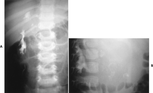 |
| FIG. 29-2 Nephroblastoma (Wilms’ tumor). A, Intravenous urogram anteroposterior projections show diffuse amorphous calcification of a large right upper quadrant mass in a pediatric patient. B, Intravenous urogram lateral view of this same patient demonstrates the retroperitoneal location of this mass. Nephroblastoma is the most common abdominal malignancy of children, with 80% occurring between 1 and 5 years of age. |
CLINICAL COMMENTS
Patients with Wilms’ tumor present with an abdominal mass in more than 90% of cases. 13 Approximately 50% of patients experience hypertension or fever. Hematuria is relatively uncommon. 13,19 No male or female predominance is noted. 13 Up to 30% with the rare condition of aniridia (congenital absence of the iris) develop nephroblastoma. 19 Four to five percent of cases show bilateral tumors. 19
• Wilms’ tumor is the most common abdominal malignancy of children.
• Abdominal mass is the most common clinical presentation.
• Radiographically, Wilms’ tumor is seen as a complex renal mass; 5% to 10% have internal calcification.
Nephrocalcinosis
BACKGROUND
Based on its location, calcification in the kidney can be classified broadly into two categories: calcification within the pyelocalyceal lumina or nephrolithiasis, and intraparenchymal calcification or nephrocalcinosis. Nephrocalcinosis also can be categorized with respect to its predominant location. A limited group of diseases, cortical nephrocalcinosis, may produce calcification confined to, or predominantly located in, the renal cortex. Other conditions, such as medullary nephrocalcinosis, spare the cortex and cause calcium salt deposition in the medulla, interstitium, or lumina of the nephrons.
IMAGING FINDINGS
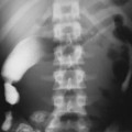
Radiographically cortical nephrocalcinosis can be differentiated from the medullary variety by the peripheral location of the calcification (Fig. 29-3). Medullary calcification is typically that of bilateral, diffuse, fan-shaped clusters of stippled calcifications, primarily in the renal pyramids (Fig. 29-4).
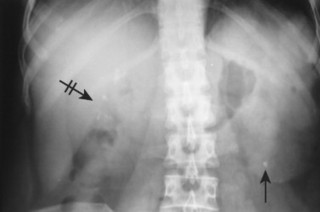 |
| FIG. 29-3 Nephrocalcinosis. Cortical (arrow) and medullary (crossed arrow) nephrocalcinosis. |
 |
| FIG. 29-4 Medullary sponge kidney. A, Faint calculi are seen in the renal pyramids (arrows). Medullary sponge kidney is characterized by ectatic collecting ducts and associated small cysts communicating with these ducts. B, Renal tubular acidosis. This parenchymal renal calcification results from renal tubular acidosis. The calcifications occur within the tubules surrounding the calyces (arrows). |
CLINICAL COMMENTS
The two most common etiologies of cortical nephrocalcinosis are acute cortical necrosis and chronic glomerulonephritis, although radiographically evident calcification is rare in both conditions. Persons on chronic dialysis may have similar calcifications.
Roughly 40% of cases of medullary nephrocalcinosis are attributable to primary hyperparathyroidism, another 20% to renal tubular acidosis, and the remaining 40% divided among many other causes.
• Renal intraparenchymal calcification is known as nephrocalcinosis.
• Two categories of nephrocalcinosis are cortical and medullary.
• Peripheral location is seen with cortical nephrocalcinosis.
• Cortical nephrocalcinosis is rare.
• Bilateral, diffuse, fan-shaped clusters of stippled calcifications typically are seen with medullary nephrocalcinosis.
• Primary hyperparathyroidism and renal tubular acidosis are the most common causes of medullary nephrocalcinosis.
BACKGROUND
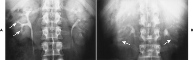
Nephrocalcinosis is calcification in renal parenchyma, whereas nephrolithiasis is concretion calcification in the luminal portion of the urinary tract (Fig. 29-5). The composition of renal stones varies with geography. In the United States, approximately 75% of renal stones are calcium oxalate. The remaining 25% of renal calculi are noncalcareous and are composed of uric acid, struvite, or cystine.
 |
| FIG. 29-5 Staghorn calculus in two different patients. A, Plain film of the abdomen demonstrates a calculus that conforms to the shape of the calyces, infundibulum, and pelvis. B, Intravenous pyelogram showing bilateral duplication of the collecting systems with a staghorn calculus obstructing the upper collecting system on the right (arrow). |
Related posts:


Stay updated, free articles. Join our Telegram channel

Full access? Get Clinical Tree


