42 2D Time-of-Flight MRA
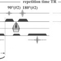
A two-dimensional (2D) spoiled gradient echo pulse sequence is typically used for 2D time-of-flight (TOF) MR angiography (MRA). Slices are acquired sequentially, in a single-slice mode, one slice at a time. A short repetition time (TR) is employed together with a high flip angle. This results in reduced signal from background (stationary) tissue, as there is insufficient time for recovery of longitudinal magnetization (T1 relaxation). On the other hand, protons moving into the slice (flow) have full longitudinal magnetization, as they have yet to be exposed to an RF pulse. The net result is that blood flowing into the slice has high signal intensity. The high signal resulting from inflow of unsaturated blood protons is referred to as flow-related enhancement and is the basic contrast mechanism in a TOF acquisition. If arterial flow is principally along one axis, and venous return in the opposite direction (such as in the neck, with flow to the brain via the carotid arteries and return via the jugular veins), saturation pulses can be used to eliminate the signal from flow in one direction. For example, to eliminate the signal from the jugular veins, an additional RF (saturation) pulse is applied superior to the axial slice. This presaturation pulse (see Case 101) moves together with the axial slice as each subsequent slice is acquired. Blood flowing in the craniocaudal direction (in the jugular veins) is thus saturated as it flows into the imaging slice and produces no MR signal. This approach was commonly used prior to the advent of contrast-enhanced MRA to image the carotid bifurcations.
Stay updated, free articles. Join our Telegram channel

Full access? Get Clinical Tree


