14 Spin Echo Imaging
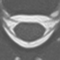
Sagittal and axial T1-weighted spin echo images, acquired at 1.5 T, are shown prior to (Fig. 14.1A,B) and axial images after (Fig. 14.1C) intravenous gadolinium chelate administration. The scans demonstrate a heterogeneously enhancing hypothalamic mass, by stereotactic biopsy, a grade II astrocytoma.
In an MR pulse sequence, either a gradient magnetic field or an RF pulse can be employed to form (refocus) the observed signal (the echo). A sequence that uses a gradient to refocus the echo is referred to as a gradient echo pulse sequence. If there is an RF pulse prior to the echo (classically a 180° pulse), then the pulse sequence is referred to as a spin echo sequence. Spin echo (SE) technique was widely used historically. However today, its application in MR is limited, largely due to the emergence of fast or turbo spin echo technique. SE technique is still commonly employed at 1.5 T for T1-weighted imaging in the brain, as illustrated in Fig. 14.1.
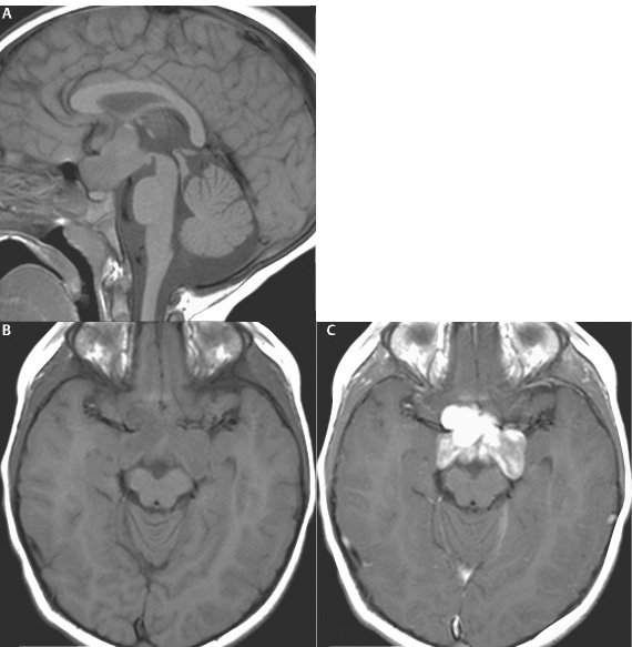
Fig. 14.1
Related posts:

Stay updated, free articles. Join our Telegram channel

Full access? Get Clinical Tree


