7 Imaging Basics: k Space, Raw Data, Image Data
The signal for MRI comes from the nuclei of hydrogen. Because the hydrogen nucleus consists of a single proton, it is common practice to refer to the signal as coming from protons. On exposing the patient to a magnetic field, more nuclear magnetic moments (of protons) will be aligned with (parallel to) the main magnetic field rather than against it. This results in a net longitudinal magnetization. Tilting of that magnetization can be done with an RF pulse. Once the magnetization is tilted away from the parallel alignment, it will start to precess around the direction of the main magnetic field with the Larmor frequency. That frequency is a function of the local magnetic field strength. To tilt the magnetization, the frequency of the RF pulse must match the rotational frequency of that magnetization. Establishing a magnetic field gradient along the direction of slice selection (e.g., z) and using an RF pulse with a limited frequency range will result in a slice-selective excitation (Fig. 7.1). In this and the figures that follow, GS is the slice-selective gradient, GP is the phase-encoding gradient, and GR is the readout gradient. Magnetizations of different resonance frequencies remain untouched. After a 90° RF pulse, the longitudinal magnetization is converted into transverse magnetization. The latter is responsible for the observed MR signal and will be encoded with spatial information. Figure 7.1 is an illustration of a slice-selective excitation using the dependency of resonance frequencies on local magnetic field strength; 1 and 2 mark the lower and upper frequency range, respectively, covered with the slice-selective RF pulse.
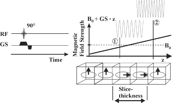
Fig. 7.1
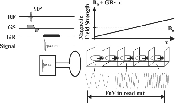
Fig. 7.2
Essential to spatial encoding in MRI is the fact that the resonance frequency of the magnetization is a function of the local magnetic field strength. Establishing a magnetic field gradient across an object (e.g., B0

Stay updated, free articles. Join our Telegram channel

Full access? Get Clinical Tree


