and Bruce J. Barron2
(1)
Loyola University Medical Center, Maywood, Illinois, USA
(2)
School of Medicine, Emory University, Atlanta, USA
1 Infections and Inflammations
1.1 Gallium-67 Citrate Scan
Indications
- 1.
Lymphoma (HD > NHL).
- 2.
Solid tumors (lung, melanoma, HCC (hepatoma), sarcoma, testicular tumor, head and neck cancers, neuroblastoma.
- 3.
Infections and inflammations (vertebral osteomyelitis – better than In-111 WBC), fever of unknown origin (FUO), fungal infections, granulomatous diseases, and sarcoidosis.
Ga-67 Citrate
t phys 78 h. Cyclotron produced, decays by electron capture (EC). Emits gamma radiation 93 (37 %), 185 (20 %), 300 (17 %), and 395 (5 %). Known as “90, 190, 290, and 390.”
Mechanisms of Action
Iron analog. Will not cross the BBB (blood brain barrier).
Infections: Ga-67 binding affinity – lactoferrin > > siderophores > transferrin. Leukocytes will secrete lactoferrin (lactoferrin mechanism), and bacteria will secrete siderophores. Ga-67 – bound to transferrin → transported via blood stream to infection site → attached to lactoferrin >> > and siderophores.
Solid tumors: Transmembrane transferrin receptor (CD71) on tumor cells via endocytosis (transferrin mechanism), then attached to lysosomal proteins.
Lymphomas: Transferrin and lactoferrin mechanism.
Protocol
Bowel prep. optional → IV injection → 48–72 h, whole body scan + SPECT/CT of the chest/abdomen at 48 or 72 h. Can wait up to 7–10 days to image (e.g., differentiate intra-abdominal infection from normal bowel clearance).
Dose
Infection (5 mCi), tumor imaging (10 mCi).
Imaging
Collimator – medium energy parallel, 20 % windows at 93, 185, and 300 KeV or 20 % windows at 93 and 185 KeV. Whole-body planar anterior posterior images followed by SPECT/CT of region of interest (chest/abdomen and pelvis).
Critical Organ
Colon.
Distribution
Blood pool (plasma protein bound %) – 24 h, 20 %; 48 h, 10 %; and 72 h, 5 %.
Infection site/tumor uptake at 12–24 h.
Liver > bone marrow >> > colon (variable uptake), lacrimal gland, nasopharyngeal, breasts (cycle variant), testes, lung, thymus (peds), spleen, kidneys.
Clearance
First 24 h – 25 % clearance by kidneys. > 24 h – bowel> > kidneys (kidney uptake is abnormal at 48 h).
Variations
- A.
Chemotherapy decreases liver activity significantly.
- B.
Scrotal uptake may be normal.
- C.
Imaging quality: Poor due to “downscatter” from high-energy photons not being imaged.
- D.
Weak bone agent. Discordance with bone scan – infection is less likely.
- E.
Sarcoidosis involving the salivary glands will demonstrate intense Ga67 uptake also known as the “panda sign”.
Distribution and Clearance

Distribution
Blood pool (plasma protein bound %) – 24 h, 20 %; 48 h, 10 %; and 72 h, 5 %. Infection site/tumor uptake at 12–24 h.
Liver > bone marrow >> > colon (variable uptake), lacrimal gland, nasopharyngeal, breasts (cycle variant), testes, lung, thymus (peds), spleen, kidneys.
Clearance (1) First 24 h – 25 % clearance by kidneys. (2) ≥ 24 h – bowel> > kidneys. (3) 48 h – 75 % tracer in body.
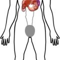
Normal Distribution
Distribution
Blood pool (plasma protein bound %) – 24 h, 20 %; 48 h, 10 %; and 72 h, 5 %. Infection site/tumor uptake at 12–24 h.
Liver > bone marrow >> > colon (variable uptake), lacrimal gland, nasopharyngeal, breasts (cycle variant), testes, lung, thymus (peds), spleen, kidneys.
Clearance (1) First 24 h – 25 % clearance by kidneys. (2) ≥ 24 h – bowel> > kidneys. (3) 48 h – 75 % tracer in body.
Abnormal Distribution