The Carotid Segments, the Aberrant ICA, and the Persistent Stapedial Artery
3.1 Case Description
3.1.1 Clinical Presentation
A 32-year-old man presented with pulsatile tinnitus and a retrotympanic vascular mass.
3.1.2 Radiologic Studies
See ▶ Fig. 3.1; ▶ Fig. 3.2.
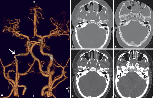
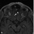
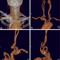
Fig. 3.1 3D CTA in anteroposterior (AP) view (a) demonstrates an aberrant course of the right ICA (white arrow), running more laterally compared with the contralateral left side. Contrast-enhanced axial CT images in bone (b,c) and soft tissue (d,e) windows reveal a vascular structure corresponding to the right ICA within the right middle ear.
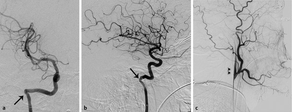
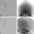
Fig. 3.2 Right ICA angiogram in AP (a) and lateral (b) views confirms the lateral course of the right ICA. The right ECA angiogram in lateral view (c) reveals the ascending pharyngeal origin of the right ICA (double arrowheads). Note the focal narrowing of the “aberrant ICA” as it enters the skull base, marking the entry point into the inferior tympanic canaliculus (arrows).
3.1.3 Diagnosis
Aberrant right internal carotid artery (ICA).
3.2 Anatomy
The aberrant ICA occurs as a hemodynamic response to a segmental agenesis of the first (i.e., cervical) segment of the ICA secondary to disturbed differentiation of the third branchial arch. It represents a collateral pathway between the external carotid artery (ECA) and the petrous ICA via the ascending pharyngeal artery (APhA).
The ICA segments have been classified by various methods, most of which are based on anatomic or surgical criteria. However, to understand the occurrence of specific variations like the one described in the present case, a model based on embryologic considerations, such as that proposed by Santoyo-Vazquez and Lasjaunias, may be more helpful. In this model, the ICA is formed by seven segments, the boundaries of which are defined by transient embryonic vessels.
As previously described, during formation of the craniocervical vessels and the arch, the ventral aorta and both dorsal aortas (DAos) are connected by six aortic arches (AAs). The lower three arches (IV–VI) participate in the development of the aortic arch and supra-aortic trunks, whereas arches I–III are involved in ICA development. Cranial to the AA I, at this stage, the DAo will also give origin to other embryonic vessels. In caudocranial order, these are the primitive maxillary artery (PMI), the dorsal ophthalmic artery (DO), and the ventral ophthalmic artery.
Bearing these considerations in mind, the future segments of the adult ICA are defined by the following embryonic vessels:
segment 1: AA III,
segment 2: the DAo between AAs III and II,
segment 3: the DAo between AAs II and I,
segment 4: the DAo between AA I and the PMI,
segment 5: the DAo between the PMI and the DO,
segment 6: the DAo between the DO and the opening of the ophthalmic artery (OA), and
segment 7: the DAo between the opening of the OA and the bifurcation of the ICA into a rostral (telencephalic) and a caudal division.
Cranially to the seventh segment, there are no additional ICA segments, as all other future arteries that will develop further distally will have a different temporal or phylogenetic origin. During embryonic life, the aortic arches and the embryonic vessels will undergo regression and significant modifications that will give shape to the ICA as it is seen in adults.
However, the remnants of these embryonic vessels will be relevant in defining the boundaries of the different ICA segments: AA III will persist and evolve into the cervical ICA distal to the carotid bulb. The carotid bulb is, from an embryological point of view, therefore a separate structure from the ICA. AA II regresses rapidly, and the only portion remaining forms the stapedial and hyoid arteries. These will be annexed by the ventral pharyngeal artery (originating from the ventral aorta) to form the future ECA and internal maxillary artery. The remnant of AA II is the caroticotympanic artery (CTymA). AA I will regress and contribute to the development of the ECA, together with the ventral aorta. The remnant of AA I will be the mandibulovidian artery. The PMI will regress to form the posterior hypophyseal artery and contribute to form the meningohypophyseal trunk. In most adults, the DO will regress and contribute to form the inferolateral trunk. The caudal division of the ICA reaches the cephalic end of the ipsilateral ventral neural artery to constitute the future PcomA. The rostral or telencephalic division will evolve initially into the anterior choroidal artery and anterior cerebral artery, which later will give rise to the middle cerebral arteries.
In cases of segmental agenesis of the ICA, each of these embryonic vessels represents the potential point of vascular reconstitution of flow into the other distally preserved ICA segments. Most of these unusual flow patterns have been improperly described as aberrant courses of the ICA. After a complex process of regression and annexation of the different vascular structures involved, the ICA develops into its final appearance, resembling a continuous vessel, with its characteristic path and curves. Still, the seven embryonic segments can be distinguished by the origin of the embryonic arteries’ remnants:
cervical segment: immediately distal to the bulb to the entry point of the of the carotid canal,

Stay updated, free articles. Join our Telegram channel

Full access? Get Clinical Tree


