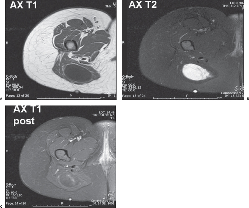
Case 30 A 27-year-old woman presents with a buttock mass. (A–C) A well-defined intragluteal ovoid lesion (L) with fluidlike signal intensity appears on axial T1- and T2-weighted magnetic resonance (MR) images. A peritumoral fat rind is visible on the T1-weighted MR image (black arrows

 Clinical Presentation
 Imaging Findings

![]()
Stay updated, free articles. Join our Telegram channel

Full access? Get Clinical Tree


