Case 30
Case History
A 56-year-old woman presents with new right breast lump.
Physical Examination
• right breast: palpable lump at 2:00 position
• left breast: normal exam
Mammogram
Mass (Fig. 30–1)
• margin: spiculated
• shape: oval
• density: equal density
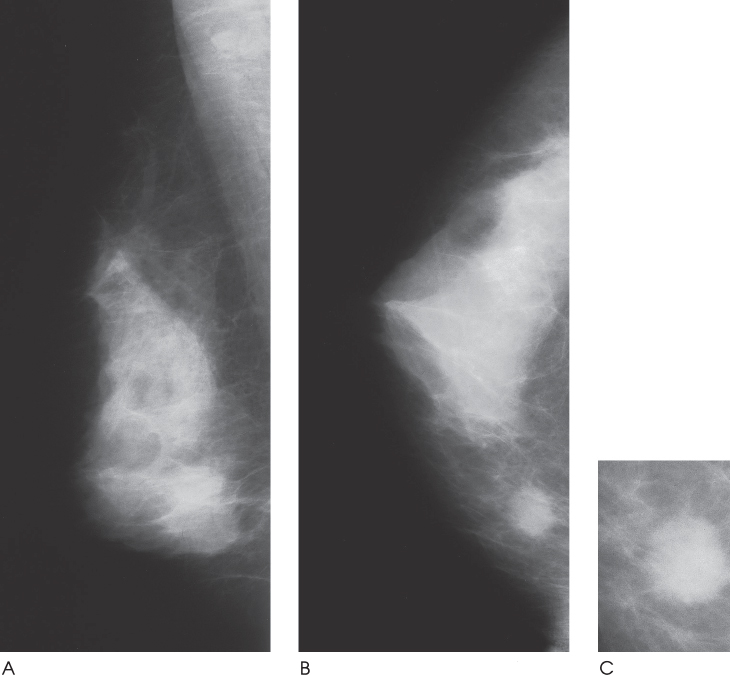
Figure 30–1. In the right upper inner breast there is an oval mass with ill-defined margins. (A). Right MLO mammogram. (B). Right CC mammogram. (C). Right CC spot compression mammogram.
Ultrasound
Low Frequency
Frequency
• 8 MHz
Mass
• margin: ill-defined


Stay updated, free articles. Join our Telegram channel

Full access? Get Clinical Tree


