Case 30
 Clinical Presentation
A 64-year-old woman with the sudden onset of flank pain and shock.
 Imaging Findings
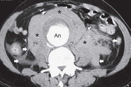
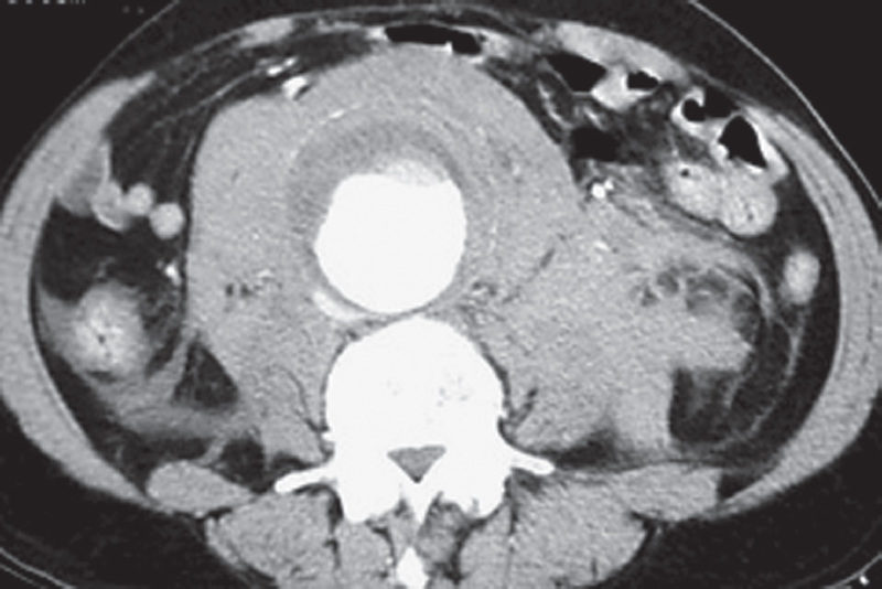
Contrast-enhanced axial computed tomography (CT) image is at the level of the middle abdomen. A large abdominal aortic aneurysm (An) is present. Surrounding the aneurysm, in the retroperitoneum, there is a hyperintense soft-tissue abnormality (asterisks) with ill-defined margins and streaks between the tissue planes (arrows).
 Differential Diagnosis
• Retroperitoneal hemorrhage from ruptured abdominal aortic aneurysm: A streaky hyperintense mass in the retroperitoneum is typical of hemorrhage in the retroperitoneum. In the presence of an abdominal aortic aneurysm, retroperitoneal hemorrhage is presumed to be due to aneurysmal rupture unless proven otherwise.
• Retroperitoneal hemorrhage from other sources: Retroperitoneal hemorrhage can occur from renal tumors, particularly renal cell carcinomas, angiomyolipomata, and adrenal neoplasms like pheochromocytomas. It can also occur spontaneously in patients with a coagulopathy. However, once an abdominal aortic aneurysm has been demonstrated, aneurysmal rupture becomes the most important diagnosis.
Stay updated, free articles. Join our Telegram channel

Full access? Get Clinical Tree


