Case 32
Case History
A 51 -year-old woman presents with a new left breast lump.
Physical Examination
• left breast: a palpable lump in the left breast at the 2:00 position
• right breast: normal exam
Mammogram
Mass (Fig. 32–1)
• margin: circumscribed
• shape: oval
• density: high
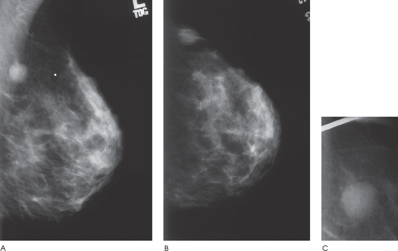
Figure 32–1. There is a well-circumscribed oval density in the left upper outer breast. This mass corresponds to a palpable lump and is new since the screening mammogram of the previous year. A small segment of the margin is indistinct even with spot compression views. (A). Left MLO mammogram. (B). Left CC mammogram. (C). Left MLO spot compression view.
Ultrasound
Frequency
• 11.5 MHz
Mass
• margin: well defined
• echogenicity: hypoechoic
• retrotumoral acoustic appearance: retrotumoral enhancement present


Stay updated, free articles. Join our Telegram channel

Full access? Get Clinical Tree


