Case 32
 Clinical Presentation
A 23-year-old woman with amenorrhea and infertility.
 Imaging Findings
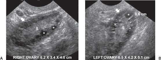
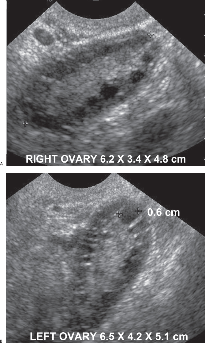
(A) Transvaginal sonographic image of the pelvis shows the right ovary (arrow), which is enlarged and has multiple peripheral small follicles in a subcapsular location (some marked with an (asterisk). There is excessive central stroma (S). No dominant follicle is seen. (B) Transvaginal sonographic image of the pelvis shows the left ovary (arrow). The findings are identical to those seen in the right ovary in Figure A (asterisks identify some of the subcapsular follicles). Central stroma (S) is excessive.
 Differential Diagnosis
• Bilateral polycystic ovaries: Enlarged ovaries with peripheral small follicles and excessive central hyperechoic stroma are characteristic.
Stay updated, free articles. Join our Telegram channel

Full access? Get Clinical Tree


