Case 33
Case History
A 68-year-old woman presents with mammographic enlargement of a left breast nodule.
Physical Examination
• normal exam
Mammogram
Mass (Fig. 33–1)
• margin: obscured
• shape: oval
• density: equal
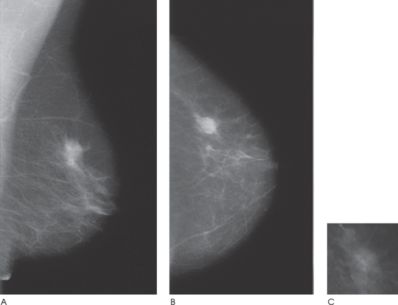
Figure 33–1. An oval circumscribed nodule with obscured margins is present in the upper outer breast. (A). Left MLO mammogram. (B). Left CC mammogram. (C). Left ML spot compression mammogram.
Ultrasound
Frequency
• 10 MHz
Mass
• margin: spiculation/architectural distortion
• echogenicity: isoechoic to fat
• retrotumoral acoustic appearance: bilateral edge shadowing


Stay updated, free articles. Join our Telegram channel

Full access? Get Clinical Tree


