Case 33
 Clinical Presentation
A 41-year-old woman with pelvic pain and excessive menstrual bleeding.
 Imaging Findings
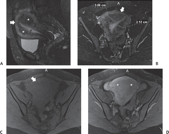
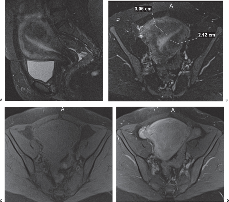
(A) Fat-saturated T2-weighted midline sagittal magnetic resonance imaging (MRI) of the pelvis shows an enlarged uterus (arrow). The bright endometrial stripe (black asterisk) is normal in thickness. However, the junctional zone (white asterisks) is diffusely but asymmetrically thickened. No punctate areas of increased signal are seen. (B) Fat-saturated T2-weighted axial MRI through midpelvic region shows the uterus (arrow), with measurements of the asymmetrically thickened junctional zone exceeding 12 mm. (C) Fat-saturated precontrast T1-weighted axial MRI through the midpelvic region shows a homogeneous appearance of the uterus (arrow) because of identical signal intensities of the junctional zone and myometrium. No punctate areas of increased signal are seen. (D)
Stay updated, free articles. Join our Telegram channel

Full access? Get Clinical Tree


