Case 34
Case History
A 59-year-old woman presents with palpable left axillary nodes. Biopsy of one of the nodes is consistent with breast cancer.
Physical Examination
• left breast: enlarged axillary node; no other palpable masses
• right breast: normal exam
Mammogram
Mass (Fig. 34–1)
• margin: circumscribed
• shape: lymph node
• density: high density
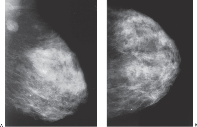
Figure 34–1. In the left axilla there are multiple enlarged axillary nodes. No suspicious mammographic masses or calcifications are present. (A). Left MLO mammogram. (B). Left CC mammogram.
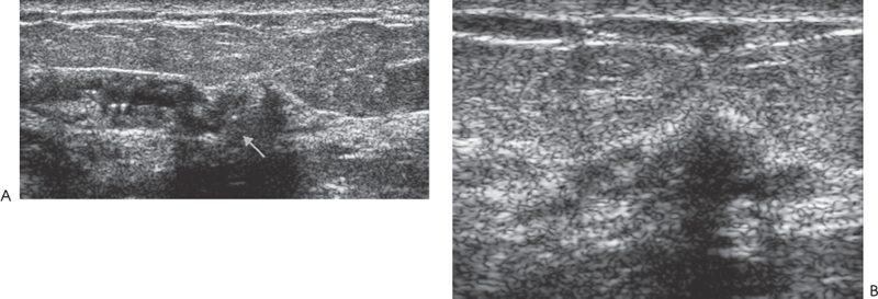
Ultrasound
Frequency
• 11.5 MHz
Mass
• margin: ill defined
• echogenicity: hypoechoic
• retrotumoral acoustic appearance: posterior shadowing distal to mass
• shape: irregular (Fig. 34–2)