Case 34

 Clinical Presentation
A 62-year-old man with testicular swelling.
 Imaging Findings

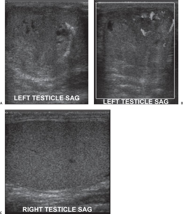
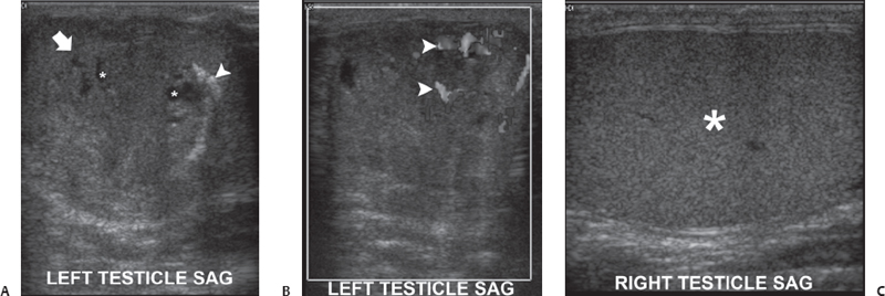
(A) Sagittal sonographic image of the left testis shows a hypoechoic, heterogeneous, ill-defined mass (arrow) within the testis. Cystic areas (asterisks) are within the mass. Calcification (arrowhead) is seen at the margin of the mass. (B) Color Doppler sonographic image of the left testis shows signal from increased internal blood flow (arrowheads) within the mass. (C) Sagittal sonographic image of the right testicle shows normal echogenicity and echotexture of the testicular parenchyma (asterisk). No focal lesion is seen.
 Differential Diagnosis
• Testicular neoplasm, most likely a nonseminomatous germ cell tumor: All solid-appearing masses of the testicle should be considered neoplastic unless there is an overwhelming history suggesting the opposite. Nonseminomatous germ cell tumors occur at the extremes of age and show a complex echotexture, with cystic and solid areas.
• Testicular hematoma:
Stay updated, free articles. Join our Telegram channel

Full access? Get Clinical Tree


