Case 35
 Clinical Presentation
A 68-year-old woman with difficulty voiding and recurrent urinary tract infections.
 Imaging Findings
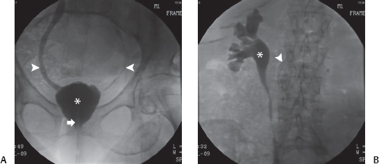
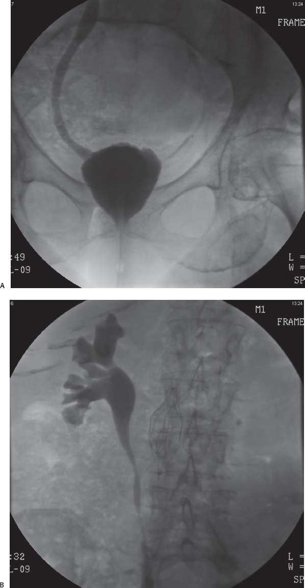
(A) Image of the pelvis during a cystogram shows the urethral catheter (arrow) in situ. The urinary bladder (asterisk) is small. Reflux of contrast into both ureters (arrowheads) is seen. The right ureter is dilated. (B) Image of the renal area during the same study shows the refluxed contrast opacifying the right collecting system (asterisk) and mild dilatation of the calices. Incidental note is made of an inferior vena cava filter (arrowhead).
 Differential Diagnosis
Stay updated, free articles. Join our Telegram channel

Full access? Get Clinical Tree


