Case 36

 Clinical Presentation
A 74-year-old man with hematuria.
 Imaging Findings

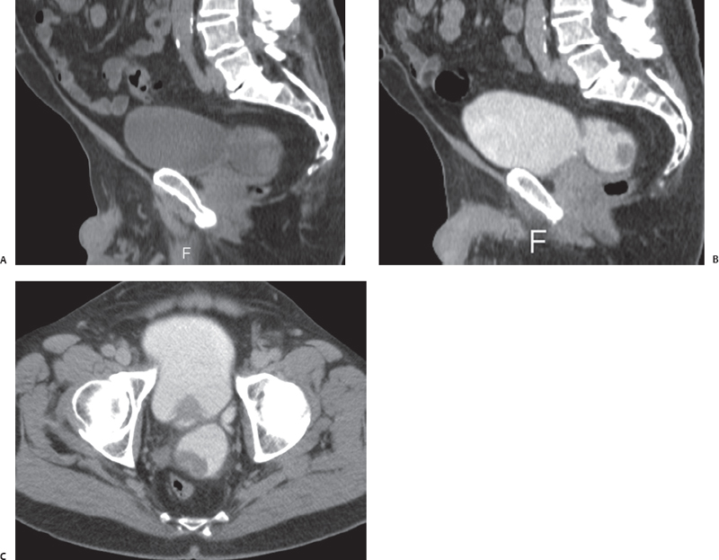
(A) Midline sagittal multiplanar reconstruction (MPR) computed tomography (CT) image of the pelvis obtained without intravenous (IV) contrast shows the urinary bladder (arrow) partially distended with urine. A diverticulum (arrowhead) arises from the posterior aspect of the urinary bladder. The prostate (asterisk) is enlarged. (B) Midline sagittal MPR CT image of the pelvis obtained in the excretory phase after IV contrast shows the urinary bladder (arrow) and the diverticulum (arrowhead) as opacified with excreted contrast. There are two filling defects in the lumen of the diverticulum (asterisks). (C) Axial source CT image of the pelvis in the same phase as Figure B shows the opacified urinary bladder (short arrow) and diverticulum (arrowhead). One of the masses in the diverticulum is also shown (white asterisk). There is retraction (long arrow) of the wall of the diverticulum where it comes in contact with the mass. The filling defect (black asterisk) in the urinary bladder is the upper portion of the enlarged prostate.
 Differential Diagnosis
• Carcinoma arising from a bladder diverticulum:
Stay updated, free articles. Join our Telegram channel

Full access? Get Clinical Tree


