Case 37
Case History
A 50-year-old woman presents with a newly discovered lump in her left breast.
Physical Examination
• left breast: palpable 1 cm mass at 12:00
• right breast: normal exam
Mammogram
Mass (Fig. 37–1)
• margin: indistinct
• shape: oval
• density: high density
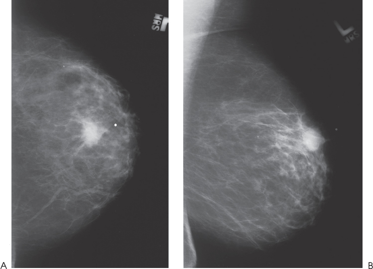
Figure 37–1. At the 12:00 position of the left breast, there is an ill-defined, oval mass that corresponds to the palpable lump. (A). Left MLO mammogram. (B). Left CC mammogram.
Ultrasound
Frequency
• 10 MHz
Mass
• margin: ill defined
• echogenicity: hypoechoic
• retrotumoral acoustic appearance: increased acoustic transmission
• shape: lobulated (Fig. 37–2)


Stay updated, free articles. Join our Telegram channel

Full access? Get Clinical Tree


