Case 37
 Clinical Presentation
A 58-year-old man with abdominal pain.
 Imaging Findings
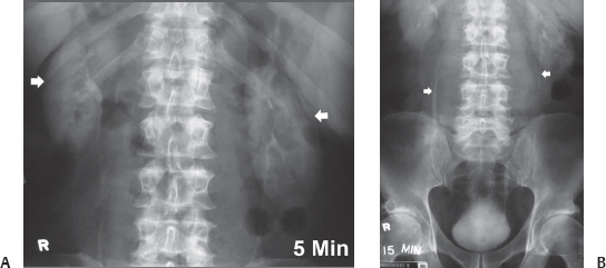
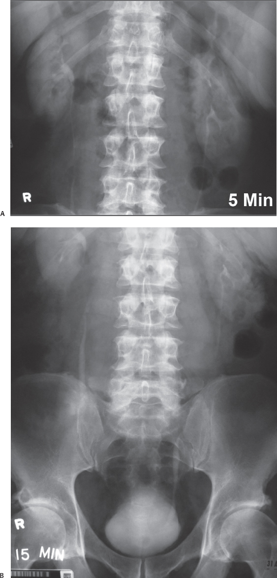
(A) A 5-minute intravenous pyelogram (IVP) image of the renal area shows that both kidneys (arrows) are normal in size, shape, position, orientation, outline, and parenchymal thickness. No dilatation of the collecting systems is seen. (B) A 15-minute full-length IVP image shows partial opacification of both ureters. The visualized portions of the ureters are normal in caliber, with no obvious filling defect. The abdominal portions of the ureters (arrows) are displaced laterally in a smooth fashion. The urinary bladder appears normal.
 Differential Diagnosis
• Retroperitoneal lymphadenopathy: This is the most common cause of symmetric lateral displacement of ureters in their abdominal course. Large lymphadenopathy can be due to lymphoma or metastatic disease.
• Retroperitoneal mass: Retroperitoneal sarcoma can displace the kidneys as well as the ureters. The displacement is usually more localized and more pronounced ipsilaterally.
• Abdominal aortic aneurysm:
Stay updated, free articles. Join our Telegram channel

Full access? Get Clinical Tree


