Case 38
 Clinical Presentation
A 62-year-old woman who recently noticed an abdominal mass while practicing yoga.
 Imaging Findings
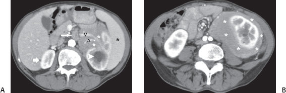
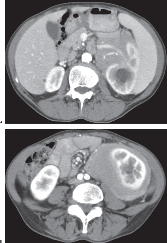
(A) Contrast-enhanced computed tomography (CT) image at the level of the kidneys shows amorphous soft tissue (white asterisks) in the left renal sinus that extends outward to encase the left renal artery (A) and vein (V) without causing significant compression. Mild dilatation of the left collecting system (arrowhead) is present. The right kidney shows a tiny simple cyst (short arrow) but is otherwise normal. A lymph node (long arrow) is seen in the retroperitoneum. The spleen (black asterisk) is enlarged. (B) Contrast-enhanced CT image at the level below that of Figure A shows amorphous soft tissue (asterisks) infiltrating the perinephric space and surrounding the kidney without causing deformity.
 Differential Diagnosis
• Lymphoma: Involvement of the perinephric region and renal sinus by amorphous soft tissue is highly suggestive of lymphoma. The absence of compression of the renal parenchyma and hilar structures is characteristic. Splenomegaly and retroperitoneal lymph node provide additional evidence supporting the diagnosis.
• Perinephric hematoma:
Stay updated, free articles. Join our Telegram channel

Full access? Get Clinical Tree


