Case 39
Case History
A 43-year-old woman presents with a palpable lump. Lump was first detected 1 year ago and had been found to be sonographically solid.
Physical Examination
• left breast: 2 to 3 cm mass at the 1:00 position
• right breast: normal exam
Mammogram
Mass (Fig. 39–1)
• margin: circumscribed
• shape: oval
• density: equal density
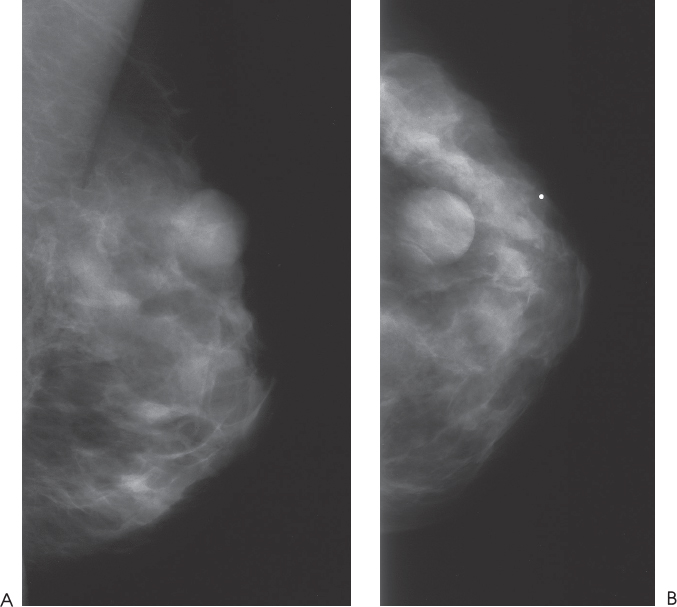
Figure 39–1. At the 1:00 position of the left breast there is a predominantly circumscribed oval mass that corresponds to the patient’s palpable lump. (A). Left MLO mammogram. (B). Left CC mammogram.
Ultrasound
Frequency
• 7.5 MHz
Mass
• margin: well defined


Stay updated, free articles. Join our Telegram channel

Full access? Get Clinical Tree


