35 3D Imaging: Basic Principles
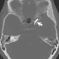
In Fig. 35.1A, a small, high signal intensity abnormality (arrow) is noted within the body of the lateral ventricle on a 2D sagittal gradient echo T1-weighted scan of the brain at 3 T acquired in 1:52 min:sec using a 4-mm slice thickness. The lesion corresponds to one of many small fat globules scattered throughout the ventricular system and subarachnoid space in this patient with a ruptured dermoid, a pathognomonic imaging presentation. This small fat globule is equally well seen on a 1-mm sagittal image (Fig. 35.1B) from a 4:21 min:sec 3D acquisition.
There are two main approaches to acquiring MR images: 2D and 3D. In a 2D acquisition, a slice is selectively excited by use of a gradient magnetic field (slice-select gradient) and then encoded in two dimensions (phase encoding and readout). The image in Fig. 35.1 A is an example of such an acquisition. In a 3D acquisition, a volume or thick slab is excited rather than a single slice. To produce slices from the slab, additional phase encodings are applied along the slice direction. The image in Fig. 35.1 B was acquired in a 3D fashion at 3 T using a low SAR fast spin echo sequence known as SPACE (see Case 37
Stay updated, free articles. Join our Telegram channel

Full access? Get Clinical Tree


