43 3D Time-of-Flight MRA
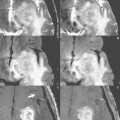
In Fig. 43.1, a 7 × 9 mm2 saccular aneurysm (arrow) is noted arising at the left middle cerebral artery (MCA) bifurcation on a maximum intensity projection (MIP) image from a 3D time-of-flight (TOF) MRA of the circle of Willis at 3 T. In Fig. 43.2, (A) an axial source image from the MRA scan is illustrated, together with (B) coronal and (C) sagittal images reformatted from the original axial acquisition, all depicting the aneurysm (A, white arrow). Note that the aneurysm incorporates multiple MCA branches and thus is not amenable to endovascular treatment (coiling).
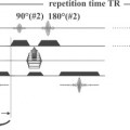
Flow is an intrinsic contrast mechanism that can be imaged with MRI in several ways. Spin echo (SE) sequences use a 90° RF pulse followed by a 180° RF pulse to produce an echo (the observed signal). In standard imaging sequences, both the 90° and 180° pulses are slice selective. In typical vessels with normal flow velocities, blood is not in the slice to receive both RF pulses, and thus flow is seen as a signal void. Gradient echo (GRE) sequences use only a single RF excitation pulse, and the echo is formed by a gradient magnetic field reversal. In this situation, blood that flows into the slice is hyperintense. This is particularly true when a short TE is used.
Stay updated, free articles. Join our Telegram channel

Full access? Get Clinical Tree


