Case 4
Case History
A 73-year-old professional violinist presents for a screening mammogram. She has a history of a soft mass in the upper outer left breast for more than 20 years.
Physical Examination
• left breast: very soft area of asymmetry in the upper outer quadrant
• right breast: normal exam
Mammogram
Mass
• margin: circumscribed
• shape: round
• density: fat-containing
Associated Findings
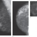
• completely fatty encapsulated mass in the upper outer quadrant of the left breast (Fig. 4–1)
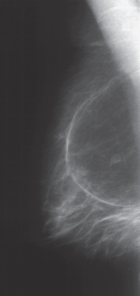
Figure 4–1. Right MLO mammogram: There is a large, well-defined radiolucent lipoma. The small nodule that projects in the center of the mass is a lymph node located just medial to the lipoma on the CC view.
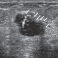
Ultrasound


Stay updated, free articles. Join our Telegram channel

Full access? Get Clinical Tree


