Case 40
Case History
A 45-year-old woman presents for screening mammogram.
Physical Examination
• normal exam
Mammogram
Mass (Fig. 40–1)
• margin: indistinct
• shape: oval
• density: high density
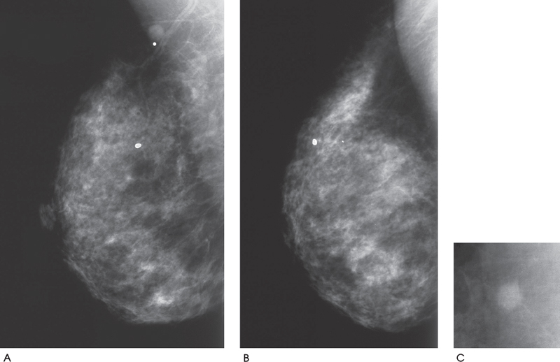
Figure 40–1. In the right upper outer quadrant, there is an ill-defined oval mass. (A). Right MLO mammogram. (B). Right exaggerated CC mammogram. (C). Right MLO spot compression mammogram.
Ultrasound
Frequency
• 13 MHz
Mass
• margin: ill defined
• echogenicity: hypoechoic


Stay updated, free articles. Join our Telegram channel

Full access? Get Clinical Tree


