Case 40
 Clinical Presentation
A 72-year-old man with diabetes underwent contrast-enhanced computed tomography for abdominal pain. The next day, he experienced backache. A radiograph of the lumbar spine was obtained.
 Imaging Findings
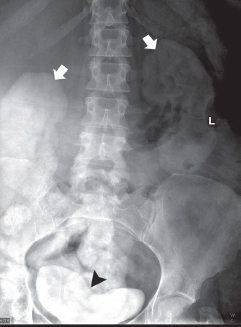
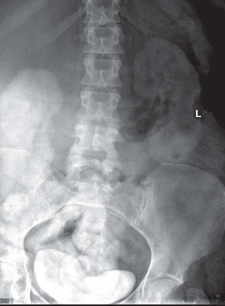
Anteroposterior view of the lumbar spine shows that both kidneys (arrows) and the urinary bladder (arrowhead) are denser than expected. Because the patient had contrast-enhanced computed tomography (CT) the day before this image, the increased density is due to persistent nephrograms. The size, shape, location, orientation, and outline of the kidneys are normal. Oral contrast administered during the CT examination is also seen and is of no clinical significance.
Stay updated, free articles. Join our Telegram channel

Full access? Get Clinical Tree


