Case 41
 Clinical Presentation
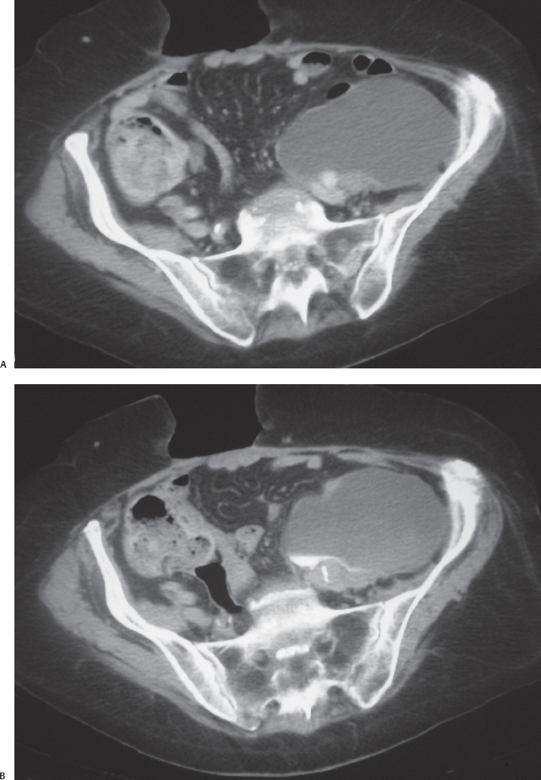
A 78-year-old man underwent descending colectomy for colonic infarction. Postoperatively, he developed fever and a fluctuant mass in the left lower quadrant.
 Imaging Findings
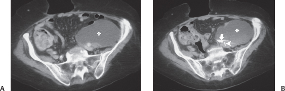
(A) Contrast-enhanced computed tomography (CT) image of pelvis shows a fluid collection in the left lower quadrant (asterisk). (B) Delayed CT image at a lower level shows excreted contrast (arrow) within the collection (asterisk). An incidental note is made of wall calcification (arrowhead) in the left common iliac artery.
 Differential Diagnosis
• Urinoma due to ureteric injury at the time of surgery:A fluid collection adjacent to the urinary tract is typical. Its opacification with contrast excreted by the urinary tract is confirmatory.
Stay updated, free articles. Join our Telegram channel

Full access? Get Clinical Tree


